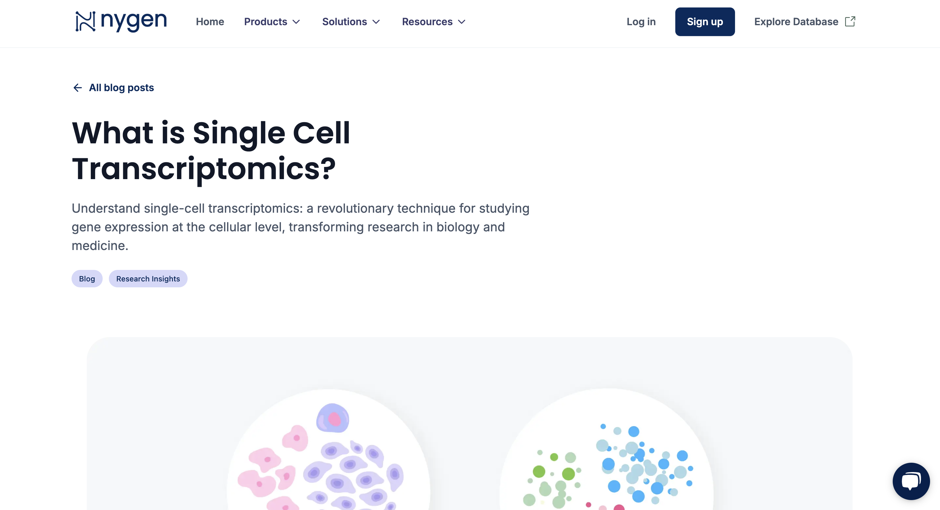
What is Single Cell Transcriptomics?
Discover single-cell transcriptomics, a transformative technique for analyzing gene expression at the cellular level in biology and medicine.
Traditional bulk RNA sequencing averages the gene expression signals across all cells in a sample. While useful, this approach can mask the variability between individual cells, particularly in heterogeneous tissues where different cell types may have distinct functions. Single-cell transcriptomics overcomes this limitation by providing data on a per-cell basis, allowing for:
- Identification of Rare Cell Types: Detecting cell populations that are present at low frequencies but may have significant biological importance.
- Understanding Cellular Heterogeneity: Revealing differences among cells that contribute to tissue function or disease states.
- Tracing Developmental Lineages: Mapping the progression of cells as they differentiate during development or in response to stimuli.
Single-cell transcriptomics involves a series of steps, each crucial for generating high-quality, interpretable data. The workflow comprises sample preparation, single-cell isolation, RNA extraction and cDNA synthesis, library preparation and sequencing, data processing, and data analysis and visualization. Understanding each of these steps in detail is essential for researchers aiming to leverage this powerful technology.
1. Sample Preparation
Successful single-cell transcriptomics starts with preparing a high-quality cell suspension. This involves tissue dissociation using enzymes like collagenase or trypsin, along with gentle mechanical disruption. Maintaining cell viability and RNA integrity is crucial, achieved through rapid processing at low temperatures and using RNase inhibitors. Cell viability assessment and filtration ensure a clean preparation of healthy cells for analysis.
2. Single-Cell Isolation
Isolating individual cells is crucial to prevent RNA cross-contamination. Various methods are used:
- Microfluidic Platforms: Systems like 10x Genomics Chromium encapsulate single cells with barcoded beads, enabling high-throughput processing for large-scale studies.
- Fluorescence-Activated Cell Sorting (FACS): Offers high purity and specificity for selecting specific cell populations, but requires specialized equipment.
- Manual Techniques: For low-throughput or specific cell isolation, methods like micromanipulation or laser capture microdissection are employed.
3. RNA Extraction and cDNA Synthesis
Due to the minimal RNA in a single cell (10-20 picograms), sensitive methods are crucial for RNA extraction and reverse transcription. Gentle lysis buffers release RNA while inhibiting RNases, with microfluidic systems often performing lysis in droplets to minimize degradation.
Reverse transcription converts RNA to cDNA using high-efficiency reverse transcriptases. Oligo(dT) primers target polyadenylated mRNA, while random hexamers capture non-polyadenylated transcripts. SMART-seq2 employs template switching for full-length cDNA enrichment and universal primer addition.
cDNA amplification is necessary for sequencing. PCR is common but can introduce biases, while linear amplification methods like In Vitro Transcription (IVT) can reduce these. Unique Molecular Identifiers (UMIs) help correct amplification biases by labeling individual RNA molecules.
4. Library Preparation and Sequencing
Library preparation for sequencing involves cDNA fragmentation, adaptor ligation, and quality control. Fragmentation occurs via mechanical or enzymatic methods. Adaptors are attached to cDNA fragments for sequencing, while indexing barcodes enable multiplexing.
PCR amplification enriches the library, followed by quality assessment using tools like the Agilent Bioanalyzer. High-throughput sequencing platforms then generate large volumes of accurate data.
5. Data Processing and Analysis
After sequencing, raw single-cell RNA-seq data must be transformed into interpretable information. The process begins with quality control to eliminate low-quality reads and artifacts, using tools like FastQC. Because single-cell data contain cell barcodes and Unique Molecular Identifiers (UMIs), specialized software such as Cell Ranger, STARsolo, or Salmon with Alevin is employed. These tools handle demultiplexing of reads by cell barcodes, align reads to a reference genome, process UMIs to remove PCR duplicates, and quantify gene expression levels for each cell. The result is a gene expression matrix where each column represents a single cell and each row represents a gene.
Normalization adjusts for technical variability like differences in sequencing depth, employing methods such as TPM or CPM. Batch effect correction is crucial when combining data from multiple sources. Software tools like Seurat and Scanpy offer comprehensive pipelines for these steps, including normalization and batch correction which requires computational understanding for application. Commercial solutions like 10x Genomics' Cell Ranger, or Partek Flow provide optimized workflows for their data.
Platforms like Nygen Analytics are designed for researchers without extensive computational expertise. Nygen simplifies data processing, making it efficient and user-friendly. It integrates these processing steps into an accessible workflow, automating tasks from quality control to normalization.
6. Data Visualization
Visualizing single-cell data is essential for interpreting complex datasets. Dimensionality reduction techniques like PCA, t-SNE, and UMAP reduce data dimensions, allowing cells to be plotted in two or three dimensions to reveal patterns and clusters. Tools like Seurat and Scanpy generate these visualizations, enabling cells to be color-coded based on metadata such as cell type or gene expression levels.
Interactive platforms like Loupe Cell Browser and BioTuring's BBrowser enhance data exploration by allowing dynamic interaction with the plots. Nygen's platform incorporates advanced visualization tools within its analytics suite, enabling researchers to create publication-quality figures without coding. Users can customize visual elements, explore gene expression patterns, and annotate cell populations interactively.
By providing an integrated solution for both data processing and visualization, Nygen streamlines the workflow from raw data to actionable insights. This approach reduces complexity and enhances accessibility, similar to comprehensive platforms like Qiagen's CLC Genomics Workbench, allowing researchers to focus on biological interpretation rather than computational challenges.
1. Validating and Refining Disease Models
When developing disease models—such as organoids, spheroids, or patient-derived xenografts—it's imperative to ensure that these models accurately mimic the cellular composition and gene expression profiles of the actual diseased tissue. Single-cell sequencing allows for a detailed comparison between the cell populations in your model and those in the native organ or tumor.
By analyzing individual cells, you can:
- Assess Model Fidelity: Determine whether your model replicates the diversity of cell types and states found in the disease context.
- Identify Aberrant Cell Populations: Detect unintended cell types or differentiation states that may compromise the model's relevance.
- Optimize Model Conditions: Adjust culture conditions or genetic modifications to better align the model with the in vivo scenario.
For instance, using single-cell transcriptomics to compare a liver organoid model with primary liver tissue can reveal differences in hepatocyte maturation or the presence of non-parenchymal cells, guiding improvements in the model.
2. Constructing Comprehensive Cellular Atlases
The scientific community is engaged in ambitious projects to map all cell types within organisms, both in healthy and diseased states. Initiatives like the Human Cell Atlas aim to create a reference map of every cell type in the human body using single-cell technologies.
Cell atlases can serve as an engine for drug discovery by revealing cell diversity and function, accelerating target validation, improving clinical trials, and inspiring new therapies.
Single-cell transcriptomics contributes to this endeavor by:
- Identifying Novel Cell Types: Discovering previously unrecognized or rare cell populations that may play crucial roles in physiology or pathology.
- Elucidating Cellular Hierarchies: Unveiling differentiation pathways and lineage relationships among cells.
- Understanding Disease Heterogeneity: Characterizing the cellular composition of tumors or inflamed tissues to identify targets for therapy.
By generating detailed cellular maps, researchers can:
- Advance Precision Medicine: Tailor treatments based on the specific cellular makeup of a patient's disease.
- Facilitate Biomarker Discovery: Identify cell-type-specific markers for diagnosis or monitoring.
- Enhance Drug Development: Target therapies to specific cell populations implicated in disease processes.
While single-cell transcriptomics offers deep, actionable insights, it also presents challenges:
- Technical Variability: Differences in cell capture efficiency, library preparation, and sequencing depth can introduce biases.
- Data Complexity: The high dimensionality of the data requires sophisticated computational tools and expertise.
- Cost: Although costs have decreased, single-cell experiments can still be expensive, particularly for large-scale studies.
- Interpretation: Translating findings into biological understanding requires careful analysis and validation.
Related articles

Integrating Multi-Omics Data for Effective Target Identification in Drug Discovery
Discover how multi-omics integration is reshaping drug discovery by uncovering disease mechanisms, prioritizing drug targets, and connecting genomics, epigenomics, transcriptomics, proteomics, and metabolomics into a usable biological model.
Read more →
Enriching Insights with Single-cell RNA-seq: Integrating Spatial Data Strategically
Discover how integrating spatial transcriptomics with scRNA-seq data enhances biological insights by mapping gene expression to tissue architecture. Learn key integration methods and real-world applications.
Read more →
Spatial Transcriptomics and Single-cell RNA-seq: Complementary Technologies for Next-Generation Biology
Discover how spatial transcriptomics and single-cell RNA-seq complement each other to drive next-generation biological insights. Learn about emerging platforms, multi-omics integration, and how Nygen Analytics empowers researchers to leverage both technologies.
Read more →